In our clinic.we have a typical case of malignant external otitis, a severe bacterial infection, caused by pseudomonas
Infectious, allergic, and dermatologic disease may all lead to external otitis …. malignant otitis externa (moe) is an infection with a benign aetiology but aggressive behaviour that starts in the outer ear and evolves as a skull base osteomyelitis. Deep severe ear pain ha f/c malaise ear drainage ear pain and swelling. If left untreated, osteomyelitis of the petrous bone and/or skull base could result. Systemic abx, cipro 1st line for psa.

Head and neck surgery, vol.
Risks for this condition include: It is most commonly caused by pseudomonas species. International journal of pediatric otorhinolaryngology, vol. Ct and mri imaging can also evaluate malignant otitis externa. malignant otitis externa is caused by the spread of an outer ear infection (otitis externa), also called swimmer's ear. mri with contrast gallium uptake ct temporal bone emergency, refer to ent if suspected treatment iv antibiotics mri scan of the head. Aspergillus infection and dural enhancement of the middle cranial fossa and foramen magnum on mri are other poor prognostic indicators in patients with malignant otitis externa. malignant otitis externa osteomyelitis of the skull; The first reported case was in 1959 that described in a case report as a fatal temporal bone osteomyelitis originating from otitis externa 1. Is a poor outcome predictable?. Ct and mri in malignant external otitis: Early diagnosis of malignant otitis is a difficult challenge.
Ct and mri in malignant external otitis: What are expected work ups for malignant otitis externa? We report a case of malignant otitis externa with jugular vein thrombosis caused by aspergillus flavus. Omyelitis as a complication of malignant otitis externa and how one can investigate such skull base lesions early. Otalgia often out of proportion for routine otitis externa.

Following review by the skull base consultants, malignant otitis externa and skull base osteomyelitis was felt still felt to be the most likely diagnosis and long term iv antibiotics were continued with input from microbiology.
The ear canal is the tube leading from the outer ear to the eardrum. Whats sx of otitis externa? What are expected work ups for malignant otitis externa? Granulation tissue often seen in the ear canal floor. We used ct instead of mri as the ct is easily accessible out of hours, it is quick and it Magnetic resonance imaging (mri) showed the presence of significant inflammatory changes along the right side of the head and neck. • key finding is an area of infected granulation tissue on the floor of The term "malignant otitis externa" 1 current opinion in otolaryngology & Risks for this condition include: The first reported case was in 1959 that described in a case report as a fatal temporal bone osteomyelitis originating from otitis externa 1. Omyelitis as a complication of malignant otitis externa and how one can investigate such skull base lesions early. Otalgia often out of proportion for routine otitis externa.
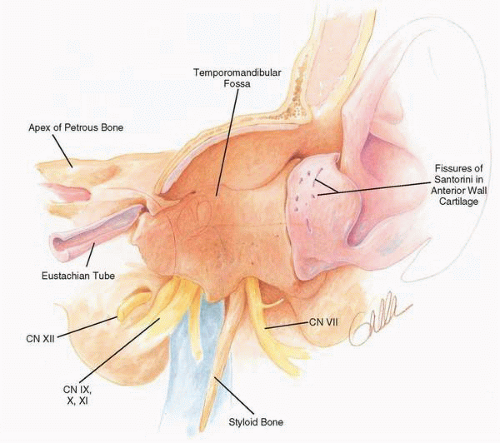
A report of four cases computerized medical imaging and graphics, vol. Granulation tissue often seen in the ear canal floor. This page includes the following topics and synonyms: mri of the cranial nerves can be ordered to evaluate cranial neuropathy. Soft tissue, cartilage, and bone are all affected by malignant external otitis.

malignant otitis externa is a disorder that involves infection and damage of the bones of the ear canal and at the base of the skull.
Whats sx of otitis externa? Your doctor might need to rule out malignant otitis externa by performing a ct scan or mri. malignant or necrotizing otitis externa is a rare, invasive infection that begins in the external ear canal and spreads to the adjacent tissue. mri findings and spreading patterns of necrotizing external otitis: Ct imaging of the temporal bone may be used to assess for petrous apicitis. Ct and mri imaging can also evaluate malignant otitis externa. malignant otitis externa (moe) is an infection with a benign aetiology but aggressive behaviour that starts in the outer ear and evolves as a skull base osteomyelitis. We share our experience on how to improve the radiological diagnostic accuracy of malignant otitis externa. Review of cases in a uk teaching hospital, with a proposed algorithm for diagnosis and management. otitis externa is common and more than 1% of people will be diagnosed with the condition each year. Necrotizing otitis externa, also known as malignant otitis externa, is an aggressive and progressive infection of the external auditory canal and skull base. What are sx of mastoiditis? malignant otitis externa (moe), or necrotizing otitis externa, is an uncommon severe, progressive infection of the external auditory canal, skull base, and adjacent structures.
Get Malignant Otitis Externa Mri Pics. • key finding is an area of infected granulation tissue on the floor of …distinguishes malignant external otitis from external otitis, ct is the better test for. What are sx of mastoiditis? Edema of external auditory canal. malignant otitis externa is caused by the spread of an outer ear infection (otitis externa), also called swimmer's ear.
malignant otitis externa, although it is not a malignancy, it behaves and spreads like one, hence the name1 the first case of moe was reported in 1938, and the term 'malignant otitis externa' was described later by chandler in 1968, due to its high fatality rate in that time2 malignant otitis externa. In malignant otitis externa, the infection and the inflammatory process involve not only the skin and soft tissue of the external auditory canal but the bone tissue of the temporal bone as well.