Nodular melanoma is an invasive form of melanoma.melanoma is a potentially dangerous skin cancer that arises from pigment cells (melanocytes).
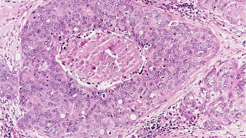
2 of 6 normal mole. Biopsy specimens from 225 benign and malignant melanocytic lesions were examined after im … Slide 118, lymph node with metastatic malignant melanoma. Doctors your own question and get educational, text answers — it's anonymous and free! (malignant b lymphocytes) accumulate in the bone marrow, interfering with the production of normal white blood cells, red blood cells, and platelets.consequently, patients may develop infections related to low white blood cell count, anemia and fatigue due to a lack of red blood cells, or easy bleeding due to a low platelet count.

malignant melanoma is one of the most common cancers in the usa, australia and europe.
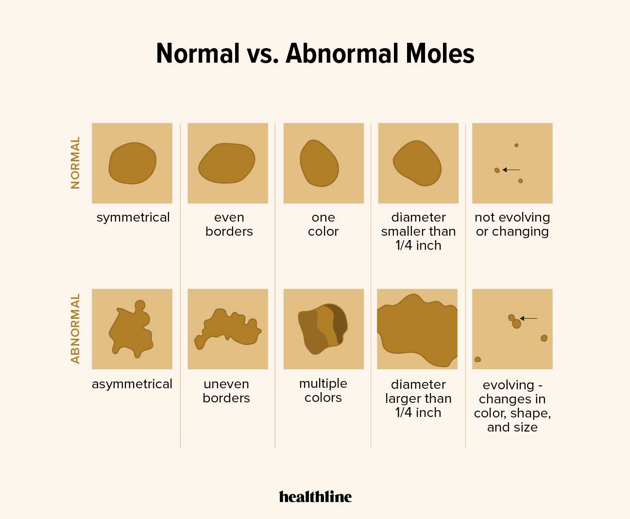
Slide 118, lymph node with metastatic malignant melanoma. Some benign moles may develop into skin cancer (melanoma). Loss of normal symmetry ! However, its changes in shape and color. Based on the diagnosis, a biopsy. 1.42 ng/mg total protein (mgp), p<0.001). Slide 151, seborrheic keratosis of skin. malignant growths often take on the same shape and color as harmless moles, so it's important to understand the difference between the two. 5 of 6 squamous cell carcinoma. Between 2004 and 2010, for example, 98% of patients diagnosed with melanoma that had not spread beyond the initial location on the skin survived 5 years or longer. As of january 1, 2018, use of the ajcc, 8th edition, 2018 is mandatory. Cuterebriasis is a parasite causing skin infections in dogs and cats. Moreover, the following symptoms can be a warning sign to you and help you to determine a normal mole from an unusual one.
Doctors your own question and get educational, text answers — it's anonymous and free! 1.42 ng/mg total protein (mgp), p<0.001). Carroll provides an accurate diagnosis based on the appearance of the lesion and the patient's clinical history. Superficial spreading melanoma is due to the development of malignant pigment cells (melanocytes) along the basal layer of the epidermis. Loss of normal symmetry !

The bottom most layer is the actively dividing basal cell layer.
Significantly higher concentrations of catl were detected in malignant melanomas than in normal surrounding skin (6.73 vs. 25 to 30% of malignant melanomas arise de novo (in previously normal skin) while the rest of the cases arise from a preexisting lesion. There are four main types of melanoma: malignant melanoma is one of the most common cancers in the usa, australia and europe. The bottom most layer is the actively dividing basal cell layer. Classifying a lesion as such is vital to your health. Moreover, the following symptoms can be a warning sign to you and help you to determine a normal mole from an unusual one. Cutaneous malignant melanoma (cmm) is a potentially lethal form of skin cancer. Most moles are benign and no treatment is necessary. This is the most common type, accounting for about 70% of melanoma cases.it can occur anywhere on the body, but is found more frequently in the upper back, legs in women and torso in men. normal moles are usually much rounder, with smooth borders. This overview of normal moles pictures includes pictures of moles and other skin spots that you can use as a first comparison to any moles on your body. For comparison's sake, 12,310 new soft tissue sarcomas are estimated to be diagnosed in 2016, while 76,380 new melanomas are expected to be diagnosed, according to the american cancer society (acs).
About 25% develop within an existing melanocytic naevus, which can be a normal common naevus, an atypical or dysplastic naevus, or a congenital naevus. Significant correlations between malignant melanoma and normal skin concentrations for catl were found. It has been estimated that 86% of melanomas can be attributed to exposure to ultraviolet (uv) radiation. Sometimes it can develop from moles. Classifying a lesion as such is vital to your health.

However, its changes in shape and color.
Based on the diagnosis, a biopsy. moles develop on nearly everybody, and are significant primarily because they can become dysplastic or malignant and need to be differentiated from melanoma. melanomas usually present as pigmented skin lesions, but they can occur on mucosal surfaces, such as in the eyes, anal. Other risk factors include inheritance, fair skin, and caucasian race. The pictures of normal moles give you an indication of what a healthy mole looks like and what characteristics it has. 4 of 6 basal cell carcinoma. 1.42 ng/mg total protein (mgp), p<0.001). However, many melanomas are new growths. Clinical types of melanoma are superficial spreading melanoma, nodular melanoma, lentigo maligna melanoma, and acral lentiginous melanoma. (malignant b lymphocytes) accumulate in the bone marrow, interfering with the production of normal white blood cells, red blood cells, and platelets.consequently, patients may develop infections related to low white blood cell count, anemia and fatigue due to a lack of red blood cells, or easy bleeding due to a low platelet count. Inflammation is another sign that a mole may be developing into a melanoma and needs to be checked out by a doctor. Sometimes it can develop from moles. This describes melanoma, the most dangerous kind, but not the most common form of skin cancer.
Download Malignant Melanoma Vs Normal Mole Pictures. According to new zealand cancer registry data, 2256 invasive melanomas were diagnosed in 2008, and at least 15% were reported as nodular melanoma. Melanin is responsible for skin and hair color. malignant growths often take on the same shape and color as harmless moles, so it's important to understand the difference between the two. Moreover, the following symptoms can be a warning sign to you and help you to determine a normal mole from an unusual one. melanomas are usually assymetrical (normal moles are usually symmetrical) b.
This overview of normal moles pictures includes pictures of moles and other skin spots that you can use as a first comparison to any moles on your body malignant melanoma mole. malignant growths often take on the same shape and color as harmless moles, so it's important to understand the difference between the two.