However, that association may be confounded by ultraviolet radiation (uv), a variable closely related to both outdoor physical activity and malignant melanoma.
In this review we shall outline the pathological characteristics of the three most common malignant skin tumours with particular emphasis on aspects which influence prognosis and management. If malignant, how deeply the tumor has penetrated the skin and whether there are. A skin neoplasm is an unusual growth on your skin. When it's diagnosed early, most people respond well to treatment. However, if the area starts to change color or develop an asymmetric growth habit, then alarm whistles should go off in your head and you should seek the expertise of a dermatologist.

skin melanoma is a relatively common human cancer with an increasing incidence trend and originates from skin melanocytes, which are neural crest derived cells.
True rectal melanoma is exceedingly rare. Satellitosis identified 1 cm from lesion, melanoma. What is a skin biopsy? In addition, five cases had available. (2008) histology of melanoma and nonmelanoma skin cancer. malignant melanoma | stratum dermatology clinics. Normal moles don't typically turn into melanoma with 70% of. Similarly, a melanoma measuring 1.04 mm thick would be recorded as 1.0 mm in the pathology report and designated as t1b for staging. Schematic showing the layers and structures of skin. Robert v rouse md rouse@stanford.edu. Slide 152, epidermal inclusion cyst of the skin. In this review we shall outline the pathological characteristics of the three most common malignant skin tumours with particular emphasis on aspects which influence prognosis and management. Teri a longacre md longacre@stanford.edu.
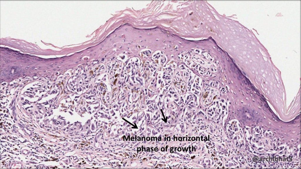
malignant melanoma in situ (mmis) is the earliest form of malignant melanoma. Ulcerated malignant melanoma, epithelioid and spindle cell types, overlying the skin of the 3rd metatarsal, clark's level iv, breslow's thickness 5.0 mm, regression not identified, ulceration present, no vascular invasion. You might also hear your doctor refer to uncertain behavior if they're not. Metastasis must be ruled out clinically or by identification of a junctional component. Is it possible not to have a concrete answer looking at the above histology report?

Slide 156, dermatofibrosarcoma protruberans of skin.
skin melanoma is a relatively common human cancer with an increasing incidence trend and originates from skin melanocytes, which are neural crest derived cells. The pathology report series is written for your patients, to help them better understand the pathologist's report. Is it possible not to have a concrete answer looking at the above histology report? However, if the area starts to change color or develop an asymmetric growth habit, then alarm whistles should go off in your head and you should seek the expertise of a dermatologist. malignant melanoma (mm) • naaman skin and laser centre. Advances in experimental medicine and biology, vol 624. Learn vocabulary, terms, and more with flashcards, games, and other study tools. None has been more emphasized and scrutinized than the histology of these melanocytic neoplasms. You might also hear your doctor refer to uncertain behavior if they're not. malignant melanoma in situ (mmis) is the earliest form of malignant melanoma. A skin neoplasm is an unusual growth on your skin. A 37 year old woman presented with a mole on the thigh which had begun to grow and change in colour over the previous few months. malignant melanoma may differ from these melanoma images.
Robert v rouse md rouse@stanford.edu. melanoma is a specific kind of skin cancer, also called malignant melanoma or cutaneous melanoma. Are expected to be diagnosed with some type of the disease. In 2020, more than 100,000 people in the u.s. melanoma is a form of skin cancer that can be lethal but can also be cured if caught very early.

Although it comprises only 3 to 5 percent of all skin cancers, it is responsible for approximately 75 percent of all.
Patient's prior shave biopsy from left dorsal forearm was reviewed in conjunction with this case. And if not what could possibly clarify it? If you are working with a site/histology that only summary stages and you need to directly code it,. melanomas typically occur in the skin but may rarely occur in the mouth, intestines or eye (uveal melanoma).in women, they most commonly occur on the legs, while in men they most commonly occur on the back. However, if the area starts to change color or develop an asymmetric growth habit, then alarm whistles should go off in your head and you should seek the expertise of a dermatologist. Persistent growth of residual, incompletely excised primary malignant melanoma, of either the epidermal or the invasive component, or both persistence or recurrence of a flat variably pigmented patch adjacent to or surrounding the scar of the primary excision site. If the physical examination shows evidence of a suspected melanoma, your doctor will recommend a skin biopsy, a procedure to remove all or part of the mole / lesion for evaluation under a microscope. Anatomical pathology there are various types of melanoma: If malignant, how deeply the tumor has penetrated the skin and whether there are. Physical activity has been positively related to malignant melanoma. The pathology report will be. The final diagnosis was nodular malignant melanoma (nm) (breslow thickness 9.5mm, clark level 4). Virtually everyone on earth has a few moles or freckles that mar their skin's surface.
Get Skin Malignant Melanoma Histology Pics. They're often categorized as benign, malignant, or precancerous. (2008) histology of melanoma and nonmelanoma skin cancer. Learn vocabulary, terms, and more with flashcards, games, and other study tools. The risk factors for skin melanoma is excessive exposure to the sun, especially in people with lighter skin. True rectal melanoma is exceedingly rare.
Ulcerated malignant melanoma, epithelioid and spindle cell types, overlying the skin of the 3rd metatarsal, clark's level iv, breslow's thickness 50 mm, regression not identified, ulceration present, no vascular invasion malignant melanoma histology. Robert v rouse md rouse@stanford.edu.


